Home » Without Label » Anatomy Rib Cage : Rib Cage Anatomy : Anterior View Of The Skeleton Of The ... / Related posts of muscle anatomy rib cage abdominal muscle diagram.
Anatomy Rib Cage : Rib Cage Anatomy : Anterior View Of The Skeleton Of The ... / Related posts of muscle anatomy rib cage abdominal muscle diagram.
Anatomy Rib Cage : Rib Cage Anatomy : Anterior View Of The Skeleton Of The ... / Related posts of muscle anatomy rib cage abdominal muscle diagram.. Ten of the twelve ribs connect to strips of hyaline cartilage on the anterior side of the body. The first rib is the widest, shortest and has the sharpest curve of all the ribs. It may occur after an obvious injury or without explanation. It consists of the 12 pairs of ribs with their costal cartilages and the sternum (figure 6.38). Anatomy of the rib cage.
The cartilage strips are called costal cartilage (costal is the anatomical adjective that refers to the rib) and connect on their other end to the sternum. Rib cage pain can be caused. See more ideas about anatomy art, anatomy drawing, anatomy sketches. The bones of the rib cage are the sternum, the 12 thoracic vertebrae and the 12 pairs of ribs. #proko #art #anatomy #ribs #ribcage #humananatomy #tutorial.
Anatomy Diagram Rib Area - Costal breathing is breathing ... from image.shutterstock.com Animated full human body anatomy. The first rib is the widest, shortest and has the sharpest curve of all the ribs. Rib cage pain can be caused. An enlarged or ruptured spleen can cause sudden or chronic pain under the left rib cage that ends up migrating towards the back and/or shoulders. The thoracic cage (rib cage) forms the thorax (chest) portion of the body. Human anatomy drawing human figure drawing anatomy study anatomy art anatomy reference figure drawing reference pose reference anatomy bones body anatomy. Two of the most notable organs behind the left side of the rib cage are the left lung and the spleen. The rib below that is rib 2, and it connects to the t2 thoracic vertebra, and so on.
The upper edge is round and the lower sharp.
The first rib is the widest, shortest and has the sharpest curve of all the ribs. The images of the human skeletal system reveal all facets of the human skeleton model (skull, spine, rib cage, shoulder, arm, hand, pelvis, leg and foot. The sternum is a flat bone that is made up of three parts, the (1) manubrium, (2) body, and the (3) xiphoid process. Ten of the twelve ribs connect to strips of hyaline cartilage on the anterior side of the body. The thoracic cage (rib cage) forms the thorax (chest) portion of the body. This furrow isn't present in the 11th and 12th ribs. Rib cage anatomy the rib cage, shaped in a mild cone shape and more flexible than most bone sets, is made up of varying elements such as the thoracic vertebra, 12 equally paired ribs, costal cartilage, and held together anteriorly by the sternum. Anatomy of the rib cage. Human anatomy drawing human figure drawing anatomy study anatomy art anatomy reference figure drawing reference pose reference anatomy bones body anatomy. Rib cage, in vertebrate anatomy, basketlike skeletal structure that forms the chest, or thorax, and is made up of the ribs and their corresponding attachments to the sternum (breastbone) and the vertebral column.the rib cage surrounds the lungs and the heart, serving as an important means of bony protection for these vital organs.in total, the rib cage consists of the 12 thoracic vertebrae and. A rib has a flat body, as you can see from the picture of the anatomy of the human rib cage. The thoracic cage protects the heart and lungs. The primary responsibilities of the ribcage involve protecting the thoracic visceral organs, enclosing the thoracic visceral organs, and is included.
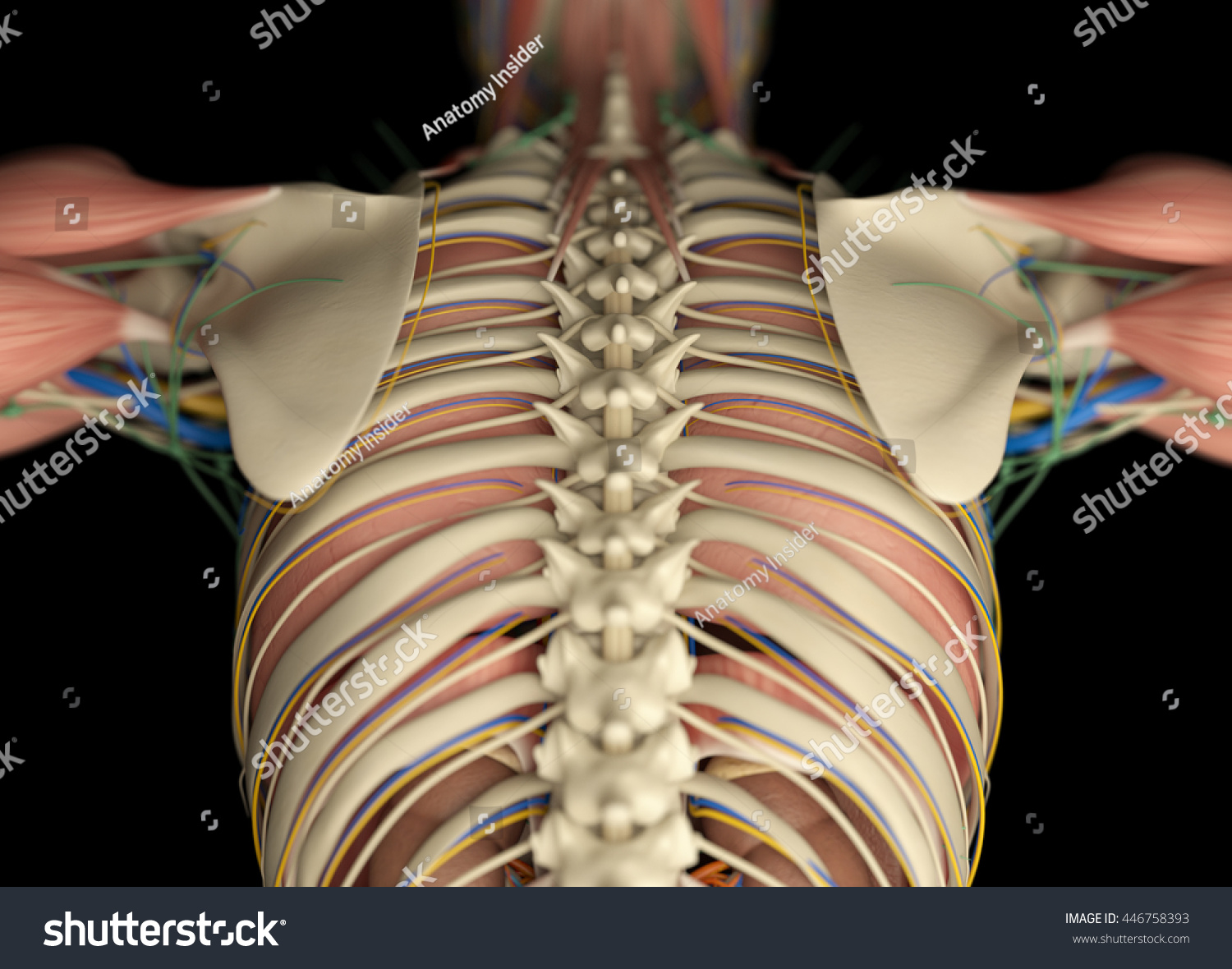
4 individual objects (spine portion, ribs, cartilages, sternum), sharing the same non overlapping uv layout map, material and pbr textures set. The head only articulates with the body of the t1 vertebra and therefore only one articulatory surface is present. Related posts of muscle anatomy rib cage abdominal muscle diagram. Anatomy of the rib cage. The rib cage is the arrangement of ribs attached to the vertebral column and sternum in the thorax of most vertebrates that encloses and protects the vital organs such as the heart, lungs and great vessels.
Ribs : Anatomy,Types,Ossification & Clinical Significance ... from howtorelief.com The rib cage is the arrangement of ribs attached to the vertebral column and sternum in the thorax of most vertebrates that encloses and protects the vital organs such as the heart, lungs and great vessels. Rib cage pain can be caused. Human anatomy drawing human figure drawing anatomy study anatomy art anatomy reference figure drawing reference pose reference anatomy bones body anatomy. The spleen is used to filter red blood cells and hangs in the upper part of the abdomen. The thoracic cage (rib cage) is the skeletal framework of the thoracic wall, which encloses the thoracic cavity. The bones of the rib cage are the sternum, the 12 thoracic vertebrae and the 12 pairs of ribs. The thoracic cage consists of the 12 thoracic vertebrae, the associated intervertebral discs, 12 pairs of ribs with their costal cartilages, and the sternum. Of all 24 ribs, the first seven pairs are often labeled as 'true.'.
Animated full human body anatomy.
Click the image to watch the anatomy of the rib cage video. An enlarged or ruptured spleen can cause sudden or chronic pain under the left rib cage that ends up migrating towards the back and/or shoulders. The lungs are responsible for processing oxygen through the body, while the spleen filters the blood and protects against some bacteria. The thoracic cage consists of the 12 thoracic vertebrae, the associated intervertebral discs, 12 pairs of ribs with their costal cartilages, and the sternum. The thoracic cage protects the heart and lungs. The spleen is used to filter red blood cells and hangs in the upper part of the abdomen. A rib has a flat body, as you can see from the picture of the anatomy of the human rib cage. Anatomy of the rib cage. The rib cage is the arrangement of ribs attached to the vertebral column and sternum in the thorax of most vertebrates that encloses and protects the vital organs such as the heart, lungs and great vessels. Each are symmetrically paired on a right and left side. The first rib is the widest, shortest and has the sharpest curve of all the ribs. Rib cage pain can be caused. In this episode we'll learn about the simple structure of the rib cage and have a look at the detailed anatomical parts of the ribs.
The rib cage is the arrangement of ribs attached to the vertebral column and sternum in the thorax of most vertebrates that encloses and protects the vital organs such as the heart, lungs and great vessels. On the interior wall of the rib body is a channel, sulcus costae, with blood vessels and nerves. The rib cage consists of 24 ribs, 12 on either side, and it shields the organs of the chest, including the heart and the lungs, from damage. #proko #art #anatomy #ribs #ribcage #humananatomy #tutorial. Rib cage, in vertebrate anatomy, basketlike skeletal structure that forms the chest, or thorax, and is made up of the ribs and their corresponding attachments to the sternum (breastbone) and the vertebral column.the rib cage surrounds the lungs and the heart, serving as an important means of bony protection for these vital organs.in total, the rib cage consists of the 12 thoracic vertebrae and.
Ribcage | Anatomy art, Skeleton drawings, Rib cage drawing from i.pinimg.com Quads only geometries (no tris/ngons). Each pair is numbered based on their attachment to the sternum, a bony process at the front of the rib cage which serves as an anchor point. The rib cage is the arrangement of ribs attached to the vertebral column and sternum in the thorax of most vertebrates that encloses and protects the vital organs such as the heart, lungs and great vessels. The primary causes of pain under the left rib cage. The lungs are two separate but connected organs located in the upper chest, covered by the rib cage. The ribs are attached to the breastbone, which is the. The first rib is the widest, shortest and has the sharpest curve of all the ribs. #proko #art #anatomy #ribs #ribcage #humananatomy #tutorial.
The thoracic cage consists of the 12 thoracic vertebrae, the associated intervertebral discs, 12 pairs of ribs with their costal cartilages, and the sternum.
Animated full human body anatomy. The thoracic cage consists of the 12 thoracic vertebrae, the associated intervertebral discs, 12 pairs of ribs with their costal cartilages, and the sternum. The thoracic cage protects the heart and lungs. Check out our anatomy rib cage selection for the very best in unique or custom, handmade pieces from our shops. The primary causes of pain under the left rib cage. The primary responsibilities of the ribcage involve protecting the thoracic visceral organs, enclosing the thoracic visceral organs, and is included. Each pair is numbered based on their attachment to the sternum, a bony process at the front of the rib cage which serves as an anchor point. The superior surface is unique in that it is marked by two grooves that allow. Related posts of muscle anatomy rib cage abdominal muscle diagram. The human rib cage is made up of 12 paired rib bones; The rib cage is the arrangement of ribs attached to the vertebral column and sternum in the thorax of most vertebrates that encloses and protects the vital organs such as the heart, lungs and great vessels. It may occur after an obvious injury or without explanation. Anatomy of the rib cage.